About MTC
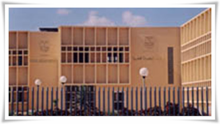
- MTC is the extension of the Medical Research Institute at Smouha district in the city of Alexandria. This center of excellency was designed to meet the challenge of doctors, researchers and scientists who are interested in the medical scientific field. This center was designed and equipped with a big number of equipment that can cope with the tremendous increase in the scientific field and the overwhelming increase in the chemical and biological analyses that have rapidly developed in last decade. This center consists of two floors; the first floor for clinical purposes and the second floor is dedicated for pure scientific research purposes. Also attached to the center a number of auxiliary services as animal house, a printing center, a guesthouse and a library.
Computed Tomography Unit ( CT-Scan ) :
- Our CT unit equipped with a CT-Scan device, general electric pro-speed. The unit is under the supervision of highly qualified professors in the filed of diagnostic imaging , following is the price list for these types of diagnostic :
- Brain
- ( SELLA ) Pituitary Gland
- Para nasal sinuses ( PNS )
- Orbits
- Petrous bone
- Lumbar spine ( ISS ) L3-s1
- Lumbar spine ( ISS ) L2-s1Â
- Lumbar spine ( ISS ) L1-s1
- Abdomen
- Abdomen & pelvis
- Pelvis
- Chest
- Pharynx
- Larynx
- Knee, shoulder, long bones
- CT sample
Electron Microscope Unit
- The Electron Microscope is a computerized Transmission Microscope, which has the following facilities:The computerized screen describes all the operating conditions of the microscope among them the magnification, the high tension, actual spot size, the focus step, the exposure time, the plate file code number and the number of images that are still available on the actual film.The magnification available is up to 200 Kev and the resolution is 0.18 nm.All types of biological tissues, in addition to, blood, bacterial and viral suspensions can be investigated using this type of computerized transmission electron microscope. For each field under investigation, a code dimensions can be established to return for more investigation and for choosing the most suitable field of interest.One of the expectations of electron microscopy is the utilization of this methodology for the diagnosis of human disease.
Immunology & Tissue Culture Unit
- The Immunology and Tissue Culture unit is equipped to serve the research and medical applications in the fields of cellular and humoral immunity and cell kinetics. It is equipped with : Biohazard cabinet; CO2 incubator; fluorescent microscope; inverted microscope; tissue homogenizers (Mechanical & Ultrasonic); ordinary and cooling centrifuges and full-automated ELISA system, Refrigerator; -20oC, -80oC, -150oC.These equipment allows the unit to work on the following applications :
- Culture of different type of cells isolated from peripheral blood, internal organs, or other tissues
- Culture of adherent cell lines
- Culture of plant cells
- Immunological assay of cellular and humoral immunity
- Functional assays for cell kinetics including cell proliferation and cytokine release
- Maintenance of cell linesStudy of virus cytopathic effectDetection of different antigens and antibodies of clinical and biological relevance
Hot laboratory for Radioisotopes
- This laboratory is devised to detect different types of hormones and tumor markers using g-counter. It also contains a b-counter, which provides highly accurate, automated counting of the level of radioactivity in radioactively tagged samples.
Atomic Absorption Unit
- Atomic Absorption Spectroscopy is today the accepted method in all fields of analysis for the determination of metallic elements. The range of application reaches from medicine and biochemistry, through agriculture and geochemistry, environmental protection and food control, to petrochemistry, metallurgy and the whole chemical industry.Main components and ultra-micro traces down to 10-9 gram can be determined with equal reliability. The double-beam principle and the computer facility give the atomic absorption, its superior base line constancy overlong measurement series. At the same time the high stability makes working near detection limit easier and produces good precision for the determination of higher concentration. In the mean time this laboratory can determine easily the following elements Cu, Zn, Na, K, Cd, Pb, Fe, V, Al and Ca in different biological fluids and in other applications in the forenamed fields. Moreover several other elements can be determined using the emission mode of the instrument.
- Gas Chromatography (GC) and High Performance Liquid Chromatography (HPLC) each have their own unique apparatus, materials and practical procedures. They are widely used for the qualitative and quantitative analysis of a large number of compounds since it has high sensitivity, reproducibility and speed of resolution.
Gas Chromatography ( GC ) :Â Gas chromatography (GC) technique is based on the partitioning of compounds between a liquid and a gas phase. It has proved to be most valuable for the separation of compounds of relatively low polarity. GC technique can be applied in the following fields:
- Clinical Chemistry: Determination of some short chain fatty acids, cholesterol and related compounds in addition to measurement of blood levels of drug metabolites and alcohols.
- Environmental Toxicology Analysis: Determination of different classes of environmental pollutants (Polycyclic aromatic hydrocarbons, aromatic amines and N-nitroso compounds). Also, can be used for determination of pesticides and drugs.
- Tests to identify and quantitate toxic drugs (clinical toxicology)
- Tests to measure trace amounts of endogenous biochemicals in body fluids.Â
High Performance Liquid Chromatography ( HPLC ): It is used for separation of a mixture of components into its constituents by passing the mobile phase, containing the mixture of the solutes, through the stationary phase, which consists of column packed with solid particles. It is the most popular form of chromatography.
HPLC technique can be applied in the following fields:
Medical Biochemistry: The options of the present instrument allow determination of a vast number of important biochemical parameters. The facility of controlling the column temperature and the presence of the auto-sampler enable the qualitative and quantitative analysis of amino acids in biological fluids. Other biochemical parameters include steroid hormones, vitamins (e.g., C, D3, A, k1, etc.), nucleotides (mono-, di- and triphosphate), etc..
Bioavailability of pharmaceutical drugs: Determination of blood level of certain drug metabolites.
Environmental Toxicology Analysis : Determination the level of some environmental pollutants.
N.B. The options of the existing instrument either the GC or the HPLC allow qualitative and quantitative analysis for other compounds more than that previously mentioned that serve in different fields of researches.The HPLC in our center can be used in the analysis of amino acids, catecholamines and many other analytes which are present in the provided catalogue of the apparatus. The price of a sample will vary according its type.
Microbiology Unit
- The unit is equipped to do research work and environmental studies as well as routine clinical microbiological work to identify microorganisms (bacterial, viral or fungal) in different clinical specimens as accurately as quickly as possible. The work in the different follows strictly the standard international rules as regarding choosing the appropriate specimen to be examined, proper sample taking to avoid contamination from normal flora and finally correct and prompt specimen transportation and storage.   Â
Routine Diagnostic Bacteriological Methods: Inoculation onto different bacteriological media at different culture conditions depends upon the type of the specimen, the site of infection and the nature of the disease to be examined.In general the final diagnosis depends upon obtaining a pure culture of the organism and identifying it by different microbiological methods and determining its sensitivity to various antibiotics.
Diagnostic Mycologic Techniques: Three approaches are applied to aid the laboratory diagnosis of fungal infection:
- Direct microscopic examination of clinical specimens and skin scraping (treated with KOH or stained with special fungal stains).
- Culture of the organism on appropriate media.(e.g. Sabouraud's agar, etc.)
- Serological tests to detect antibodies in patient's serum or spinal fluid to diagnose systemic mycosis.
Diagnostic Virology Methods: Four main approaches are used to diagnose viral diseases in clinical specimens, some of them are used in research work only and others are applied in routine diagnosis of viral infections in department, these are:
- Identification in cell culture either by a characteristic cytopathic effect (CPE) or by the use of several immunologic techniques if the virus dose not produce a CPE, such as hemadsorption, complement fixation, hemagglutination, neutralization, enzyme-linked immunosorbent assay, immunofluorescence, etc.
- Direct microscopic identification by (1) light microscope to reveal characteristic inclusion bodies or multinucleated giant cells, (2) UV microscopy is used for fluorescent-antibody staining of the virus in infected cells, and (3) electron microscopy to detect virus particles which can be characterized by their size and morphology.
- Serologic procedures to detect a rise in specific antibody titer or the presence of IgM antibody. Several immunological techniques are used routinely such as ELISA, Immunofluorescence, Western blot and RIBA techniques.
- Molecular Biology Methods. e.g. PCR technique which is used routinely to diagnose HCV and HBV.
Environmental Studies: ( TO STUDY NOSOCOMIAL AND COMMUNITY INFECTIONS )Â
Activities Done By The Microbiology Unit: The microbiology unit performs different training courses in the following fields:
- Applying typing methods for epidemiological investigation.
- Studying hospital acquired infections.
- Studying water hygiene (ultra-filtration system).
- Applying standard international methods to analyze and investigate sterile products manufactured in Egypt.  Â
- Standard microbiological methods.
- Enzyme-linked Immunosorbent Assay (ELISA).
- Basic molecular biology techniques.
- Polymerase Chain Reaction (PCR).
- Environmental examination.
Molecular Biology Unit
- Several innovative molecular biology techniques are used accurately and precisely in research work as well as in some routine diagnostic tests. Our team is highly trained and has a good experience to do the following techniques:
- Isolation and purification of total bacterial DNA.
- Isolation of bacterial plasmid (Plasmid profile).
- Electrophoresis of nucleic acids (Agarose Gel System).
- Bacterial transformation.
- Southern and Western blotting.
- Cloning techniques.
- Polymerase Chain Reaction (PCR): This molecular biology technique is used to amplify a target DNA or RNA (viral or bacterial). It can be used in the following:
- Biological research: it has accelerated the study of gene function, gene mapping and their link to genetic diseases.
- Clinical applications: include identifying viruses (HCV, HBV, HIV), bacteria (TB) and cancerous ells in human tissues.
- Forensic science: PCR created detailed fingerprinting that can definitively identify individuals.
Parasitology Unit
- Parasitology and Medical Malacology Unit aims to improve the quality of researches in the field of prevention and control of parasitic diseases especially fasciolosis and schistosomosis in the surrounding communities in the vicinity of Alexandria, Egypt and Middle East. The unit offers counseling, technical support, training and parasite bio-products for researchers from Egypt and the different countries.
- The unit maintains experimentally the life cycles of both Fasciola spp. and Schistosoma mansoni to supply the researchers with the different stages of those two parasites. Also, the unit offers a variety of antigens of the two parasites upon request.
Clinical Chemistry Laboratory
- The Clinical Chemistry Tests comprise over one-third of all hospital laboratory investigations and is involved in research into the biochemical basis of disease and in clinical trials of new drugs.
- Our clinical chemistry laboratory provides the core analyses, commonly requested tests which are of value in many patients, on a frequent basis.
Histopathology unit
- Our histopathology and cytopathology unit is fully equipped in a way that allows us to:
- Examine tissue specimens received from surgical theater (Human) or experimental animals under the light microscope.
- Examine the cytological constituents of any fluid (either urine, sputum, ascitic or pleural fluid) under the light microscope.
- Give a very rapid diagnosis while the surgeon is in the theater, by frozen sections of the tissue and be examined under the microscope.
- Stain the tissue immunologically with many tumour markers for a good accurate diagnosis. Â
Epidemiology Unit
- Epidemiology has been defined as the study of the distribution and determinants of health related states or events in specified populations, and the applications of this study to control health problems. This emphasizes that epidmiologists are concerned not only with death, illness and disability, but also with more positive health states and with the means to improve health.
- This emphasizes the important role that the unit will have in satisfying the objective of the center, and that is to establish a reference regional center for tropical disease and related non-communicable diseases of major health impact in the region. Training courses in the following topics will be arranged :
- Descriptive epidemiology.
- Clinical epidemiology.
- Analytical epidemiology.
- Clinical trials.
- Case control studies.
- Chronic diseases registrations.
- Medical research design.
- Assessment of validity of diagnostic and screening tests.Â
AUXILIARY SERVICES
LIBRARY: ( In Hadara & Smouha )
- The library contains a considerable collection of biomedical journals and books.
- Internet connection and Document Delivery Service are also available.
- The library services are available to all researchers as well as undergraduates.
- Medline searching.
- Printing result of Medline search.
- Internet.
- Sending or receiving e-mails through our account.
- Document Delivery : Collecting scientific articles from any other library inside or outside Egypt is an available service provided to any user either MRI staff or not.
PRINTING CENTER :( In Hadara )
ANIMAL HOUSE : ( In Hadara & Smouha )
- A modern equipped printing center is one of the achievements of the Egyptian-Italian Cooperation Project. It already started its activities serving Alexandria and surrounding areas. It includes most developed printing machines performing a prominent role in publishing some local scientific journals and researches. This printing and editing center is expected to offer more useful services for promoting the publishing movement in Alexandria.
NAMRI – team : ( Nanotechnology from research to application )
- This unit has been provided with proper environment, housing and care that allow small experimental animals (rats and mice) to grow, mature and reproduce and to remain in good health. The unit is provided with facilities for fish and snails.This unit will provide laboratory animals needed for research activities carried on in the Center as well as external researches with reasonable prices depending on the strain.Â
NAMRI – team vision:
- Nanotechnology laboratory Team in Medical Technology Center (MTC), Medical Research Institute (MRI), Alexandria University.
NAMRI – team mission:
- NAMRI – team does his best to be the leader in the nanotechnology and nonmaterial research to provide new discoveries and applications in this field for solving important health and environmental problems.
- NAMRI – team works in implementation of almost of the new applications of nanotechnology with different nonomaterials in health and environmental research and encourage the researchers to pursue their research activities in this field to investigate almost of all of the key areas of nanotechnology applications .
NAMRI – team currant activities:
- In the Field of Environmental Application of Nanotechnology. A current project entitled:
Impact of Nanotechnology in water and Wastewater Treatment - In the field of biomedical application:
Hyperthermia for cancer treatment using metal oxide nanoparticles,
- Green synthesis of nanoparticles using some plant extract.
- We have devices such as:
-
Electrospinning device for nanofiber fabrication.
Drug delivery using nanoparticles in combination with ionotophoresis,
Imaging using nanoparticles as contrast agent,
Antimicrobial activity of some types of nanoparticles  such as silver and zinc oxide.
Nanovaccination using nanoparticles as  adjuvant.
Nanofiber for wound dressing nanoparticles.
Rotary evaporator for preparation of liposomes, magnetoliposomes and niosomes.
A unit for iron oxide preparation.
Microwave and ultrasound for hyperthermia and enhancement drug release.
Lasers for phototherapy applications.
UV-vis spectrophotometer.
ADSL Service (2MB)


